
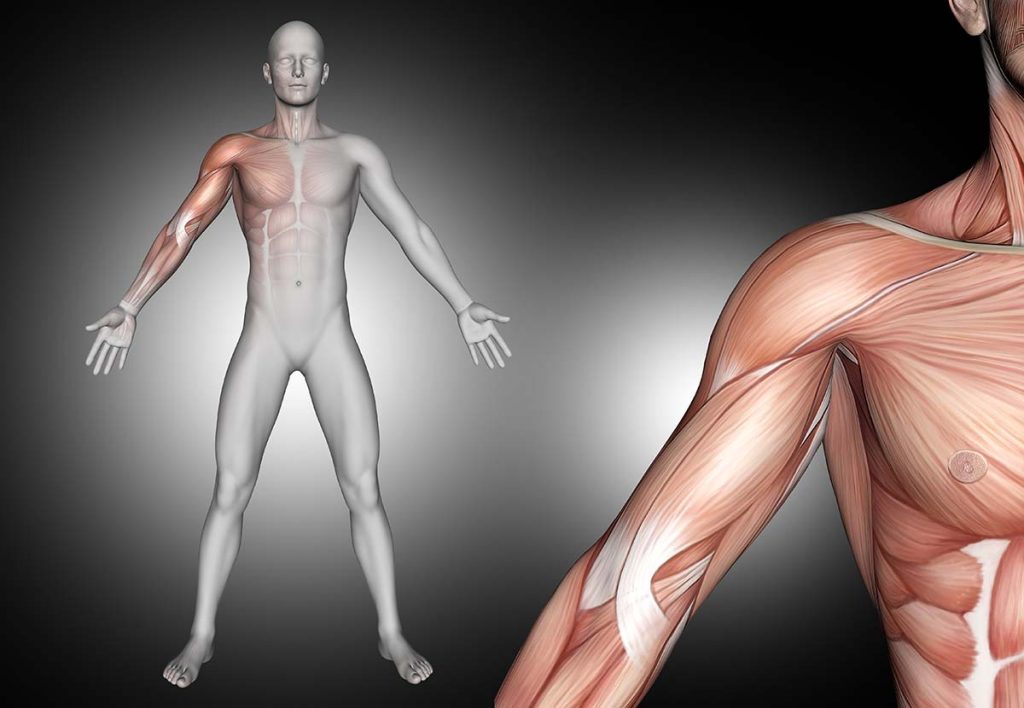
The rotator cuff muscles anatomy and functional unit are located in the upper extremities. It is a group of four muscles that hold the upper arm to the shoulder.
It provides strength and stability during arm and shoulder movement. The head of the upper arm bone, also called the humerus, fits into the scapula or the socket of the scapula.
When you extend your arm away from your body, the rotator cuff muscles prevent it from slipping out of the joint socket.
They are also called SITS muscles, referring to the first letter of their name.
The rotator cuff injury is very serious. It is common, especially among people over 40, athletes, and people who need to repeatedly raise their arms above their heads at work. Conservative treatment is usually successful.
In this article, we will learn about the anatomy of rotator cuff muscles with the clinical aspects of rotator cuff muscles.
Rotator cuff muscles anatomy
The rotator cuff muscles are a group of four muscles. For memorizing the muscles name we can retain the sentence “Rotator cuff SITS on the shoulder”. SITS means-
- S means- Supraspinatus
- I means- Infraspinatus
- T means- Teres minor
- S means- Subscapularis
These are the four muscles which are collectively called rotator cuff muscles.
Supraspinatus
Origin
The supraspinatus muscle originates from the supraspinatus fossa. A shallow depression in the body of the scapula above the spine.
Insertion
It is inserted into the greater tubercle of the humerus.
Nerve supply
The nerve supply of the supraspinatus muscle is the Suprascapular nerve, C5 & C6, the superior trunk of the brachial plexus.
Blood supply
The blood supply is the Suprascapular artery.
Action
Extend the arm from 0 degrees to 15 degrees, when it is the main active muscle. It helps the deltoid muscle to abduct to 90 degrees in this range.
Function
The main function of the supraspinatus muscle is stabilization. The action related to the supraspinatus muscle is the abduction of the humerus, which may have a weak effect on the lateral rotation of the humerus. The supraspinatus muscle, the most frequently torn rotator cuff muscle, has been the subject of extensive research.
Test of Supraspinatus
The assessment of this muscle is carried out by the Jobe’s test or commonly referred to as the “empty can” test. It’s through with a 90 degrees abduction and internal rotation (thumb pointing to the floor) of the arm while pressing down on the arm. If it is painful or weak, it is positive.
Clinical aspects
Among the four muscles of the rotator cuff, the supraspinatus muscle is the most commonly injured because it receives the greatest strength. Aging can also cause shoulder problems due to inactivity, natural atrophy of muscles and ligaments, and lifelong listening to Taio Cruz.
And Statistics show that Supraspinatus tendinitis (painful arc syndrome) is a very common disease. It is the most common inflammatory problem encountered around the shoulder joint as well as supraspinatus muscle. Typically it is seen in people aged 25-60.
Infraspinatus
Origin
The infraspinatus fossa of the scapula, with some fibers arising from the infraspinatus fossa which covers the muscle and separates it from teres major muscle and teres minor muscle.
Insertion
The posterior aspect of the greater tubercle of the humerus, and the capsule of the shoulder joint.
Nerve supply
Suprascapular Nerve (C5 & C6).
Blood supply
Suprascapular and circumflex scapular arteries.
Action
External rotation of the arm at the glenohumeral joint; stability of the humeral head in the glenoid. And helps to produce shoulder extension.
Function
It provides the main muscle strength for external rotation of the shoulder, and together with the rest of the rotator cuff muscles, it provides stability for the shoulder complex.
Test of Infraspinatus
The assessment of this muscle is performed by rotating laterally against resistance with the elbow flexed and the arm in a neutral abduction/adduction position. If it is painful or weak, it is positive.
Clinical aspects
Infraspinatus tendon tears often occur in athletes on the head due to overuse injuries or chronic shoulder instability. The main complaints are pain during sleep, weakness in the affected arm, and inability to move the arm during certain movements.
On the other hand, due to repetitive stress or normal aging, a partial tear may damage the tendon, but it will not tear completely. A full-thickness or full-thickness tear separates the infraspinatus muscle from the bone. It is usually caused by an acute injury, such as a fall.
| Read More Articles What bones make up the shoulder do you know? What are the causes & treatment of pain in my back between shoulder blades? |
Teres minor
Origin
The upper two-thirds of the lateral edge of the scapula.
Insertion
The superior fiber terminates in a tendon inserted to the lower facet of the greater tubercle of the humerus. The inferior fiber is inserted into the humerus just below the inferior facet of the greater tubercle of the humerus.
Nerve supply
Axillary nerves of posterior brachial plexus (C5 and C6 roots).
Blood supply
Circumflex scapular artery and posterior circumflex artery.
Action
- Assist shoulder adduction and extension.
- The teres minor and the infraspinatus mainly cause external rotation of the shoulder joint.
- When the humerus is stable, it abducts the inferior angle of the scapula.
Function
Together with the other muscles of the rotator cuff, the teres minor muscle provides stability to the shoulder joint and helps to keep the humeral head in the glenoid of the scapula.
Test of Teres minor
The evaluation of this muscle is carried out through a hornblower’s test, done with the arm abducted 90 degrees, the elbow bend (90 degrees), and rotate laterally against resistance. If it is painful or weak, it is positive.
Clinical aspects
Damage to this muscle may be the result of trauma, such as a fall, direct impact on the shoulder, or rapid use of force (such as pulling). The injury may also be the result of long-term overuse (repetitive head extensions, extended arms pushing or lifting) or muscle weakness.
Tendinitis is an inflammation of the tendon, a common condition caused by the overuse of the muscle. Tendon tears may also occur, which may be caused by trauma such as a fall, or they may degenerate due to long-term weakness.
Degenerative tears are more prone to degenerative tears when the muscles are not used or remain stretched for a long time (for example, sitting with round shoulders). Since the muscles also act to stabilize and compress the humeral head in the joint socket, weakness can also lead to secondary problems, including shoulder impingement.
Subscapularis
Origin
It arises from the subscapular fossa.
Insertion
Inserts on the lesser tubercle of the humerus.
Nerve supply
The upper and lower subscapular nerves (C5-C6) are innervated by the posterior brachial plexus.
The subscapularis muscles are innervated by the upper and lower subscapular nerves from the posterior cord of the brachial plexus. The upper part of the subscapularis muscle and the subscapular nerve is divided into two branches. The upper subscapular nerve supplies the upper part of the subscapularis and the lower subscapular nerve supplies the lower part of the subscapularis.
Blood supply
Subscapular artery.
Action
The primary function is the internal rotation of the humerus. Help in the adduction and extension of the shoulder in certain positions.
Arm position features a marked effect on the actions caused by this muscle: when the arm is lifted, subscapularis pulls the humerus forward and downward; when the humerus is in a fixed position, the insertion of subscapularis can act as an origin and it produces abduction of the inferior border of the scapula.
Function
Subscapularis muscle plays an important role in the stabilization of the shoulder.
Test of Subscapularis
The “take-off” and “bear hug” tests are used to evaluate this muscle.
In the “take-off/Lift-off” test, the patient takes the hands around the back to the lumbar area with the palms confronting outward. If the patient cannot lift his hands from the back, the test is positive.
In the “bear hug” test, the palm of the affected side is placed on the opposite shoulder, the fingers are stretched, and the elbows are in front of the body. The patient is then asked to maintain this posture while the examiner attempts to use external rotation to pull the patient’s hand from the shoulder to the forearm. The test result is positive when the patient cannot resist the examiner and appears weak compared to the contralateral side.
Clinical aspects
More commonly, subscapular muscle dysfunction in the form of inhibition and weakness can lead to biomechanical abnormalities of the glenohumeral joint, such as poor anterior stability of the shoulder joint in a sports shoulder.
When the subscapularis muscles become tight, weak, and/or dysfunctional, it can cause many problems: Loss of movement in the shoulders. Shoulder pain (diffuse and severe) during exercise. Shoulder weakness/loss of stability.
Key facts
| Muscles Name | Origin | Insertion | Primary function |
| Supraspinatus Muscle | Supraspinatous fossa of scapula | Greater tubercle of Humerus | Abduction of the arm to 15° at the glenohumeral joint; stabilization of humeral head in the glenoid cavity. |
| Infraspinatus Muscle | Infraspinatous fossa of scapula | Greater tubercle of Humerus | External rotation of the arm at the glenohumeral joint; stabilization of the humeral head in the glenoid cavity. |
| Teres minor Muscle | The lateral border of the scapula | Greater tubercle of Humerus | External rotation and adduction of the arm at the glenohumeral joint; stabilization of the humeral head in the glenoid cavity. |
| Subscapularis Muscle | Medial two-third of the subscapular fossa | Lesser tubercle of the Humerus | Internal rotation of arm; stabilization of humeral head in the glenoid cavity. |
Common injuries of rotator cuff muscles
Sometimes shoulder pain appears for no reason. Wear and tear of the rotator cuff may occur due to repetitive stress and neglect of posture. When this happens, the different structures around the rotator cuff may be affected.
If your rotator cuff is injured, you may feel pain or weakness when you raise your arm. Your rotator cuff injury may cause difficulty in basic functional activities (such as lifting weights, stretching, or sleeping).
Potential injuries and problems of these four rotator cuff muscles may include:
Tendinopathy
Shoulder tendinopathy refers to any injury to the shoulder tendons, whether chronic or acute. Typically due to overuse, this type of injury happened. Two types of tendon problems can occur in the shoulder: tendinitis and tendinopathy. Rotator cuff tendinitis is considered to be the mildest form of rotator cuff injury. It can be developed from the following aspects:
- Age related degeneration
- Repetitive motion
- Overuse
- Trauma
Impingement
Shoulder impingement syndrome is the result of a vicious circle of rotator cuff friction between the rotator cuff and the upper outer edge of the shoulder.
Friction can cause more swelling and further shrinkage of space, which can lead to pain and irritation.
Treatment includes rest, ice compresses, anti-inflammatory drugs, physical therapy, cortisone injections, and surgery.
Shoulder impingement syndrome usually improves within a few weeks or months, especially with the right type of shoulder exercise, but occasionally problems can occur.
Bursitis
Bursitis occurs when the bursa (a small fluid-filled sac that protects the rotator cuff) is irritated. This happens when you repeat the same actions over and over again, such as throwing a baseball or lifting things above your head. It may also be due to infection.
Common symptoms of rotator cuff muscles injury
The most common symptoms of a rotator cuff muscles anatomy injury include:
- Pain worsens at night, making it difficult for the affected side to fall asleep
- Pain when raising and lowering arms or certain movements
- Weakness when raising or rotating arms
- Creeping or cracking sensation when moving shoulders in certain positions
Some people with rotator cuff injuries may not feel any pain. This situation can be progressive, and degradation will occur slowly. According to one study, only one-third of rotator cuff tears cause pain.
Video presentation of rotator cuff muscles anatomy
FAQs of rotator cuff muscles anatomy
What causes rotator cuff injury?
“Injury” and “degeneration” are the two main causes of rotator cuff tears. Rotator cuff injuries, such as tears, can occur suddenly when falling on an outstretched hand. It will also evolve over time due to repetitive activities. Rotator cuff tears can also occur due to aging and tissue degradation.
Which rotator cuff muscle is injured most often?
Tendon tears in these muscles are called rotator cuff tears. The most commonly affected muscle of the rotator cuff muscle is the supraspinatus muscle.
When should consult with your doctor?
If your pain gets worse or starts to interfere with your normal activities or your ability to sleep well, and if you cannot use your shoulder as before, it is best to see a doctor for diagnosis and treatment. Early treatment of rotator cuff injuries can prevent increased pain and the inability to use the arms and shoulders in daily activities.
What happens if the torn rotator cuff is not repaired?
Without any treatment, whether it is rest, rehabilitation, or surgery, rotator cuff disease will get worse. Over time, you may feel more pain. You may lose the range and strength of your shoulders, making it difficult for you to perform daily activities.
Resources
- Roh MS, Wang VM, April EW, Pollock RG, Bigliani LU, Flatow EL. Anterior and posterior musculotendinous anatomy of the supraspinatus. J Shoulder Elbow Surg. 2000 Sep-Oct;9(5):436-40. [PubMed]
- Fallon J, Blevins FT, Vogel K, Trotter J. Functional morphology of the supraspinatus tendon. J Orthop Res. 2002 Sep;20(5):920-6. [PubMed]
- McCausland C, Sawyer E, Eovaldi BJ, Varacallo M. StatPearls [Internet]. StatPearls Publishing; Treasure Island (FL): Aug 10, 2020. Anatomy, Shoulder and Upper Limb, Shoulder Muscles. [PubMed]
- Valasek P, Theis S, DeLaurier A, Hints Y, Luke GN, Otto AM, Minchin J, He L, Christ B, Brooks G, Sang H, Evans DJ, Logan M, Huang R, Patel K. Cellular and molecular investigations into the development of the pectoral girdle. Dev Biol. 2011 Sep 01;357(1):108-16. [PubMed]
- Warmbrunn MV, de Bakker BS, Hagoort J, Alefs-de Bakker PB, Oostra RJ. Hitherto unknown detailed muscle anatomy in an 8-week-old embryo. J Anat. 2018 Aug;233(2):243-254. [PMC free article] [PubMed]
- de la Garza O, Lierse W, Steiner D. Anatomical study of the blood supply in the human shoulder region. Acta Anat (Basel). 1992;145(4):412-5. [PubMed]
- Moser TP, Cardinal É, Bureau NJ, Guillin R, Lanneville P, Grabs D. The aponeurotic expansion of the supraspinatus tendon: anatomy and prevalence in a series of 150 shoulders MRIs. Skeletal Radiol. 2015 Feb;44(2):223-31. [PubMed]
- Abbott, L. C., and Saunders, L. B. de C. M.: Acute traumatic dislocation of the tendon of the long head of biceps brachii; report of 6 cases with operative findings. Surgery 6: 817–840 (Dec.) 1939.
- Baer, W. S.: Operative treatment of subdeltoid bursitis. Bull. Johns Hopkins Hosp. 18: 282–284, 1907.
- Codman, E. A.: The Shoulder. Boston, Thomas Todd Co., 1934.
- DePalma, A. F.: Surgery of the Shoulder. Philadelphia, J. B. Lippincott Co., 1950.
- McLaughlin, H. L.: Lesions of the musculotendinous cuff of the shoulder; observations on pathology, course, and treatment of calcific deposits. Ann. Surg. 124: 354, 1946. [PMC free article] [PubMed]
- https://en.wikipedia.org/wiki/Rotator_cuff
- https://www.physio-pedia.com/Rotator_Cuff
- https://www.ncbi.nlm.nih.gov/books/NBK537202/
- https://www.ncbi.nlm.nih.gov/books/NBK441844/
1 thought on “Rotator cuff muscles anatomy with the details of rotator cuff”